The power of 3D imaging for improving oral surgery
The integration of three-dimensional imaging systems into oral and maxillofacial surgery practices has elevated patient care. With the ability to diagnose oral health conditions with greater accuracy than ever before, oral surgeons can now more easily identify issues and create precise surgical plans.
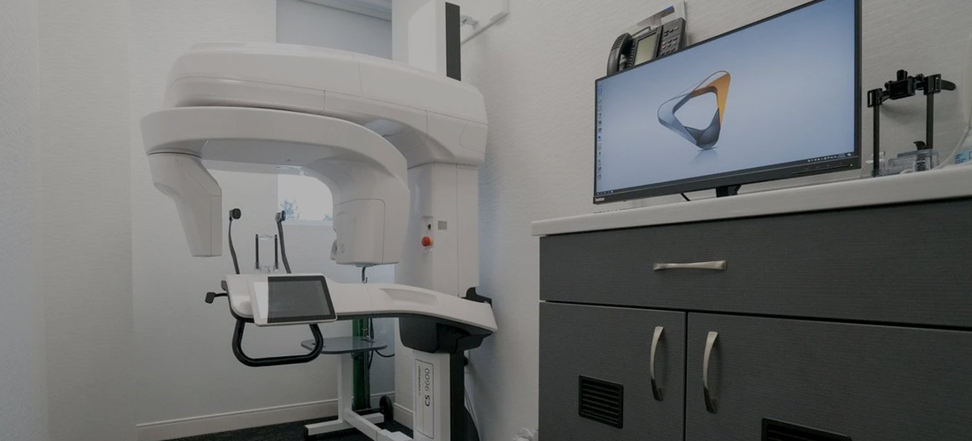
What is a CBCT digital imaging system?
CBCT, which stands for cone beam computed tomography, uses diverging x-rays in the shape of a cone to produce a 3D image. The system works in a similar fashion to a CT scanner, where the patient sits still as the CBCT machine rotates fully around their head. During the process, up to 200 images are taken from different angles. Then, all this data is compiled into a single three-dimensional model.
What can a CBCT image display?
CBCT images can show all the elements of your head and neck, including:
Bone
Soft tissues
Nerves
Roots of teeth
Cavities
Infections
Advantages for oral surgery
CBCT scans utilize less than 10% of the radiation of traditional medical imaging techniques and because the system is fast the process is more comfortable for the patient. Additional advantages of 3D imaging include:
Detailed images make it easier to diagnose complex conditions
Early detection of a wide range of oral health concerns
More accurate measurements of jaw dimensions
Dental implants and 3D imagery
CBCT scans are particularly valuable when planning complex tooth extractions or dental implant placement. 3D imagery makes it possible to more precisely determine implant size and identify the ideal placement position in relation to nerves, sinuses and adjacent tooth roots.
Posted by
boms on
Sep 23rd, 2023 11:09 pm
Filed under
Oral Surgery Blog for Patients, Referring Doctors . You can follow any responses to this entry through the
RSS 2.0 feed.
Both comments and pings are currently closed.
Authorization and Release of Testimonial Information
I understand my photo or video testimonial (the “Testimonial”) made on behalf of Bergen Oral & Maxillofacial Surgery (hereinafter called “The Practice”) may be used in connection with publicizing and promoting The Practice. I authorize The Practice to use my name, brief biographical information, and the Testimonial as defined on this form.
I hereby irrevocably authorize The Practice to copy, exhibit, publish or distribute the Testimonial for purposes of publicizing The Practice’s services or for any other lawful purpose. These statements may be used in printed publications, multimedia presentations, on websites, social media channels or in any other distribution media. I agree that I will make no monetary or other claim against The Practice for the use of the statement.
In addition, I waive any right to inspect or approve the finished product, including written copy, wherein my testimonial appears.
I hereby hold harmless and release The Practice from all claims, demands and causes of action which I, my heirs, representatives, executors, administrators or any other persons acting on my behalf or on behalf of my estate have or may have by reason of this authorization.
Data Privacy
PBHS and its Third Party Providers will have the right to collect, extract, compile, synthesize, and analyze data and information resulting from or relating to the use or operation of the services under this Agreement (“Service Data”). Any Service Data collected by PBHS or any Third Party Provider will be owned by the party collecting the Service Data and may be used by that party for any lawful business purpose without a duty of accounting to you subject to the then current privacy policy applicable to the services under this Agreement. You consent to the use and disclosure of personally identifiable and other data and information as described in this Agreement and in the then-current privacy policy applicable to the services provided under this Agreement (“Privacy Policy”).
Indemnification
You agree to and hereby indemnify, defend, and hold harmless PBHS, its Third Party Providers and their respective affiliates, employees, agents, contractors, assigns, licensees, and successors in interest (“Indemnified Parties”) from any and all claims, losses, liabilities, damages, fees, expenses, and costs (including attorneys’ fees, court costs, damage awards, and settlement amounts) that result from or relate to any claim or allegation against any Indemnified Party arising from you accessing or using the services provided under this Agreement (including any Third Party Services) or from any email or other communication generated or sent through such services or any content contained therein, whether or not in breach of this Agreement.
Messaging Program
PBHS offers subscription text message programs. Customers may ask their agent to enroll them in a text message alert program. Once enrolled in SMS texting services, you will have an opportunity to confirm or decline the service via a reply to an initial text message you receive. By agreeing to these terms of service, you are confirming a subscription to this text message program.Message frequency varies.
PBHS does not have a separate charge for this service; however,message and data rates may applyfrom your mobile carrier. Subject to the terms and conditions of your mobile carrier, you may receive text messages sent to your mobile phone. Participation in the programs on this short code is standard rated.
By providing your consent to participate in this program, you approve any such charges from your mobile carrier. Charges for text messages may appear on your mobile phone bill or be deducted from your prepaid balance. PBHS reserves the right to terminate this SMS service, in whole or in part, at any time without notice. The information in any message may be subject to certain time lags and/or delays. You are responsible for managing the types of SMS texts you receive.
Privacy Contact Information
If you have any questions, concerns, or comments about our privacy policy you may contact us using the information below:
We reserve the right to make changes to this policy. Any changes to this policy will be posted.
REQUEST AN APPOINTMENT
The first step towards a beautiful, healthy smile is to schedule an appointment. Please contact our office by phone or complete the appointment request form below. Our scheduling coordinator will contact you to confirm your appointment.
